Glaucoma is a progressive eye disease in pets that causes a dangerous rise in intraocular pressure, damaging the optic nerve and leading to blindness if untreated. Early detection can save sight, but owners often miss subtle cues. This guide walks you through the signs, diagnostic tools, and both medical and surgical treatment pathways so you can act fast when your dog or cat shows trouble.
Understanding Glaucoma and Its Impact on Pets
Glaucoma isn’t a single condition; it’s a symptom complex. The intraocular pressure (IOP) rises when fluid (aqueous humour) can’t drain properly, pushing on the optic nerve, the visual highway that sends images to the brain. When pressure stays high, the nerve fibers degenerate, and vision deteriorates.
Both dogs and cats can develop primary (genetic) or secondary (injury, inflammation, tumor) glaucoma. Certain breeds-such as the Basset Hound, Cocker Spaniel, and Siamese-carry a higher genetic load, making breed awareness a key part of prevention.
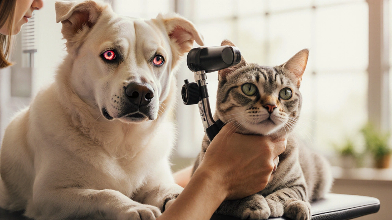
Recognizing the Early Warning Signs
Pets mask discomfort, so you need to watch for subtle changes. Common red flags include:
- Sudden squinting or keeping one eye partially closed.
- Cloudy or milky appearance of the cornea (known as corneal edema).
- Redness of the white part of the eye (sclera) that doesn’t improve.
- Frequent pawing at the eye or head shaking.
- Visible pain when you gently touch the area around the eye.
- Changes in pupil size-often a dilated, non-reactive pupil.
Because these symptoms overlap with other eye conditions, a professional evaluation is essential.
Getting a Proper Diagnosis
A veterinarian with ophthalmic training will run a series of tests:
- Tonometry: Measures intraocular pressure using a handheld device. Normal IOP in dogs and cats ranges from 10‑20 mmHg; values above 25 mmHg raise alarm.
- Gonioscopy: Examines the drainage angle of the eye to differentiate primary vs secondary glaucoma.
- Ophthalmoscopy: Checks the optic nerve head for cupping-a hallmark of chronic pressure damage.
- Ultrasound (if the cornea is cloudy) to evaluate internal structures.
Collecting baseline data helps track disease progression and guide treatment decisions.
Medical Management Options
When the disease is caught early, topical and oral medications can lower IOP and buy time.
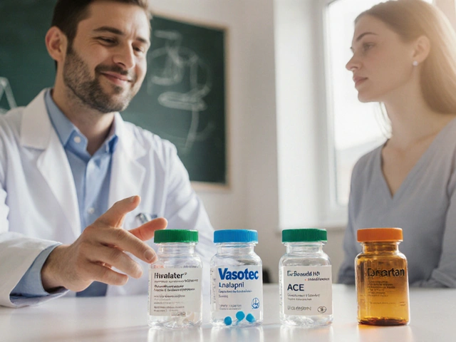
Key drug classes include:
- Carbonic anhydrase inhibitors (e.g., dorzolamide) reduce fluid production.
- Beta‑blockers (e.g., timolol) also curb fluid creation.
- Prostaglandin analogues (e.g., latanoprost) increase outflow.
- Systemic carbonic anhydrase inhibitors (acetazolamide) for severe cases.
Compliance is crucial-missed drops can cause pressure spikes within hours. Owners should set reminders and keep a log.

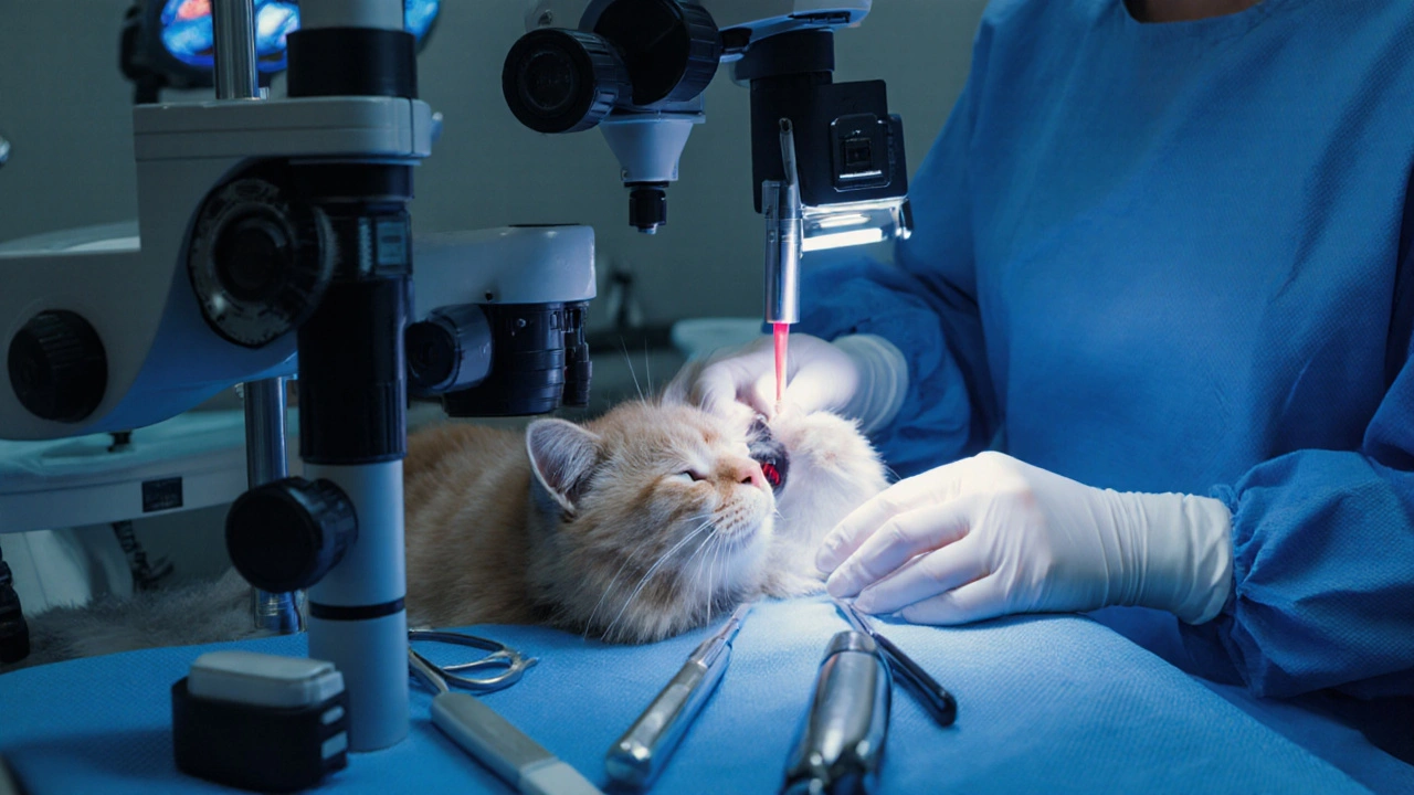
Surgical Interventions When Meds Aren’t Enough
When medication fails to keep IOP under control, surgery becomes the next line of defense. The choice depends on the animal’s age, breed, and overall health.
| Aspect | Medical Management | Surgical Management |
|---|---|---|
| Goal | Lower IOP with drugs | Alter drainage anatomy permanently |
| Typical Cost (USD) | $150‑$300 per month | $1,200‑$3,500 (one‑time) |
| Success Rate | 60‑80% short‑term | 70‑90% long‑term |
| Invasiveness | Non‑invasive (topical/oral) | Invasive (laser, cyclophotocoagulation, shunt implantation) |
| Recovery Time | Immediate (no downtime) | 1‑2 weeks with post‑op meds |
Common procedures include:
- Laser cyclophotocoagulation: Destroys part of the ciliary body to reduce fluid production.
- Implantable drainage devices (e.g., Ahmed or Molteno shunts) create a new outflow pathway.
- Iridectomy: Removes a slice of the iris to open the drainage angle.
Post‑operative care mirrors medical management-regular IOP checks and anti‑inflammatory drops are standard.
Home Care and Lifestyle Adjustments
Even with treatment, day‑to‑day habits can influence outcomes:
- Keep the environment calm; stress can raise IOP.
- Avoid exposure to bright sunlight or harsh wind, which can aggravate corneal edema.
- Maintain a healthy weight; obesity is linked to higher IOP in some breeds.
- Schedule routine veterinary eye exams every 6‑12 months for chronic cases.
Use a soft, lint‑free cloth to gently wipe away discharge-never rub the eye.
When to Seek Emergency Help
Acute angle‑closure glaucoma can develop in minutes. If you notice sudden, severe eye pain, a visibly bulging eye, or the animal refuses to eat or drink, head straight to an emergency veterinary clinic. Prompt IOP‑lowering therapy can prevent irreversible loss.
Related Topics to Explore Next
Understanding glaucoma fits into a broader pet‑health picture. You might also want to read about:
- Canine cataract prevention and treatment
- Feline retinal detachment signs
- Genetic screening for inherited eye diseases
- Nutrition’s role in ocular health
Frequently Asked Questions
Can glaucoma be cured in pets?
Glaucoma is typically a chronic condition. While you can control intraocular pressure and preserve vision, the underlying disease usually cannot be fully eradicated. Early detection and consistent treatment offer the best chance of maintaining sight.
How often should I have my pet’s eye pressure checked?
For a pet diagnosed with glaucoma, monthly checks are common during the first few months, then every 2‑3 months once stable. If your animal is at risk (breed‑predisposed), schedule a baseline exam and repeat annually.
Are there side‑effects from glaucoma eye drops?
Yes, some pets may develop mild eye irritation, conjunctival redness, or temporary blurry vision. Systemic absorption can lead to low blood pressure or heart rate changes, especially with beta‑blockers. Always report any unusual behavior to your vet.
What is the cost difference between medication and surgery?
Medication typically costs $150‑$300 per month, adding up over years. Surgical options are a one‑time expense ranging from $1,200‑$3,500, plus post‑op meds. Long‑term, surgery often becomes more economical if meds fail.
My cat’s eye looks cloudy-could it be glaucoma?
Cloudiness can signal corneal edema from high pressure, but it also appears with ulcers or infections. A veterinary eye exam with tonometry is the only way to confirm glaucoma.
Do certain diets help prevent glaucoma?
There’s no direct diet‑glaucoma link, but antioxidants (vitaminE, lutein) support overall eye health. Omega‑3 fatty acids may reduce inflammation, which can be beneficial for secondary glaucoma forms.
Can I use human glaucoma drops on my dog?
Never. Formulations differ in concentration and preservatives. Only veterinary‑prescribed eye drops are safe for pets.


Nadia Stallaert
September 27, 2025Ever wondered why the pet‑care industry seems to keep the truth about eye disease shrouded in secrecy? They whisper in sterile corridors, promising miracles while the real cost is hidden in the fine print. The very devices that measure intraocular pressure are calibrated to a narrow standard that only benefits the manufacturers. When a Basset Hound shows early signs, you’re handed a bottle of drops that cost more than a monthly electric bill, and a vet who smiles while the animal suffers. The pharmaceutical giants push carbonic anhydrase inhibitors as if they’re the end‑all, yet they ignore the root cause: genetic predisposition that they never mention in the brochure. You’ll hear about “surgical options” yet the pamphlet never speaks of the long‑term inflammation that follows a shunt implantation. Lens‑focused marketing campaigns flood social media with glossy before‑and‑after photos, but the shadows behind those images are full of pets whose vision never truly returns. In reality, the only thing that truly “cures” glaucoma is a permanent blindness that forces owners to accept a life‑long expense. The agenda? Keep the cash flowing, keep the owners dependent, keep the industry thriving. I’ve seen vets discreetly hand out “discount” cards that are, in fact, a subscription to a never‑ending supply of medication. The legal jargon in those consent forms is designed to drown out any doubt, making you sign away the right to question the procedure. And while we’re busy polishing the surgical tools, the simple act of monitoring eye pressure at home gets dismissed as “too cumbersome” for the average person. The truth is that regular home tonometry could catch spikes early, but no one sells a cheap, reliable home kit because it would undercut the lucrative clinic visits. So, before you trust the glossy brochure, remember that every “solution” comes with a hidden price tag, not just in dollars but in the silent loss of sight that many pets endure. Keep your eyes open, ask for the raw data, and demand transparency before you let your companion walk into the dark.